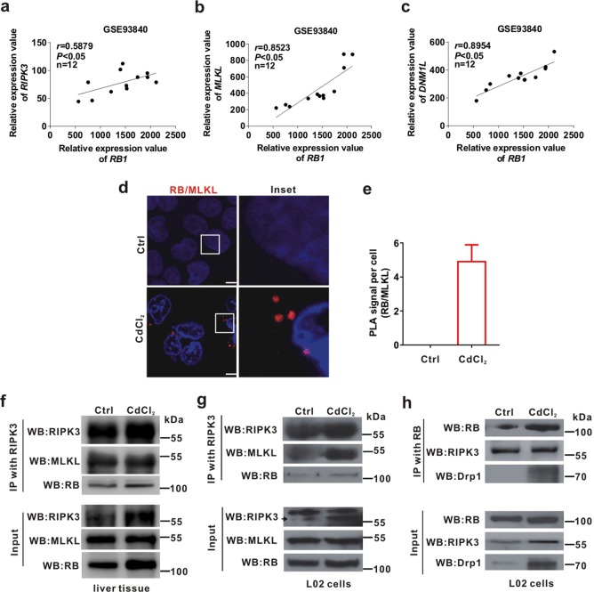
Fig. 4. RB binds to Drp1 and promotes the formation of necrosome.
a-c The linear correlation of the expression of RB1 with RIPK3, MLKL, and DNM1L expression in primary human hepatocytes exposed to three xenobiotics (aflatoxin B1, amiodarone, and chlorpromazine) for 14 days from NCBI, GEO database (Accession No. GSE93840) was calculated. d, e PLA was used for detecting interaction between RB and MLKL. Nuclei were stained with 4',6-diamidino-2-phenylindole (DAPI). The scale bar is 10 μm. d Typical red foci of PLA indicated the protein-protein interaction between RB and MLKL. e Quantification of PLA foci per cell was shown in bar charts quantified using ImagePro-plus 6.0. Ten fields of view were calculated for each group. f The ICR mice (n = 3) were intraperitoneally injected with CdCl2 (1 mg/kg bw) or physiological saline (Ctrl group, n = 3) for 7 days. The mitochondrial fraction of liver was immunoprecipitated with anti-RIPK3 antibody, and then both IP and whole cell lysates (Input) were immunoblotted with the indicated antibodies. g, h L02 cells were treated with CdCl2 (20 μM) for 6 h. The whole cell lysates were immunoprecipitated with anti-RIPK3 or anti-RB antibody, and then both IP and whole cell lysates (Input) were immunoblotted with the indicated antibodies. Data are expressed as the mean ± standard deviation (SD). P < 0.05, *significantly different from Ctrl

