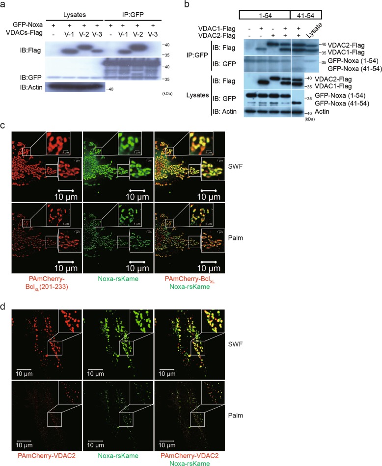
Fig. 3. MTD of NOXA binds VDAC2 protein.
a 293 HEK cells were cotransfected with GFP-Noxa (1-54) and VDAC1, 2, or 3-Flag expression plasmid vectors, and immunoprecipitation assay was carried out using anti-GFP antibody. Immunoblots were done using anti-Flag, anti-GFP, and anti-actin antibodies. b The same experiments as described in Fig. 3a were carried out except for cotransfection of expression vectors GFP-Noxa (1-54) or GFP-Noxa (41-54) plus VDAC1 or 2-Flag. c, d HeLa cells were cotransfected with PAmCherry-BclXL (201-233) or (and) Noxa-rsKame (c), and PAmCherry-VDAC2 or (and) Noxa-rsKame (d). The images for Noxa-rsKame (green), PAmCherry-BclXL (201–233) (the mitochondrial outer membrane marker; red), and PAmCherry-VDAC2 (red) were obtained by PALM and SWF. (SWF) summed widefield total internal reflection microscopy (TIRF) (total internal reflection microscope)

