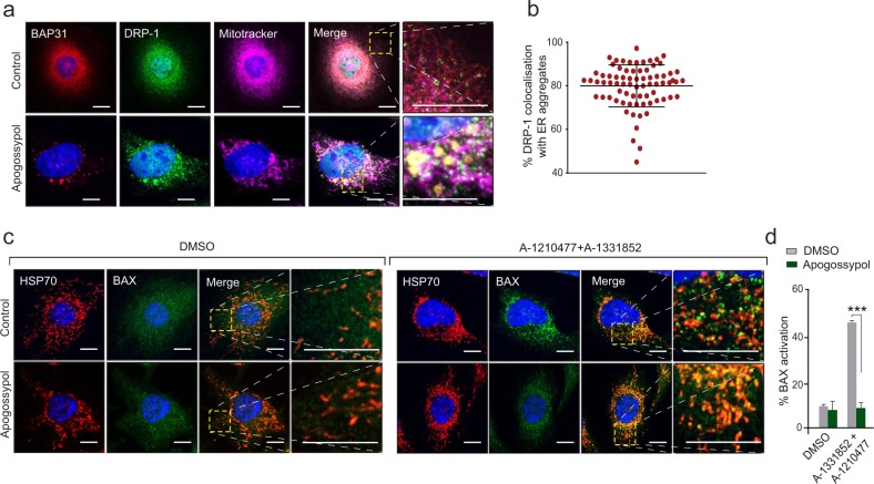
Fig. 3. Reorganised ER membranes co-localise with DRP-1 and prevent activation of BAX.
a HeLa cells were exposed to apogossypol (20 μΜ) for 4 h, stained with Mitotracker and immunostained with DRP-1 and BAP31 antibodies. b The extent of DRP-1 that was colocalised to the BAP31-positive reorganised ER tubules was assessed in 80 cells and plotted. Each dot in the graph corresponds to one cell. Error bars = Mean ± SD. c HeLa cells were exposed to Z-VAD.fmk (30 μΜ) for 30 min and apogossypol (20 μΜ) for 1 h, followed by a combination of BH3 mimetics, A-1331852 (0.1 μΜ) and A-1210477 (10 μΜ) for 4 h, and immunostained with BAX and HSP70 antibodies. Scale bar: 10 μm. In both a, c, the boxed regions in the images are enlarged to show the extent of colocalisation. d HeLa cells treated as in c were stained with BAX (6A7) antibody and the extent of BAX activation assessed by flow cytometry. Graphs were plotted using data from three independent experiments. Error bars = Mean ± SEM. ***p ≤ 0.001

