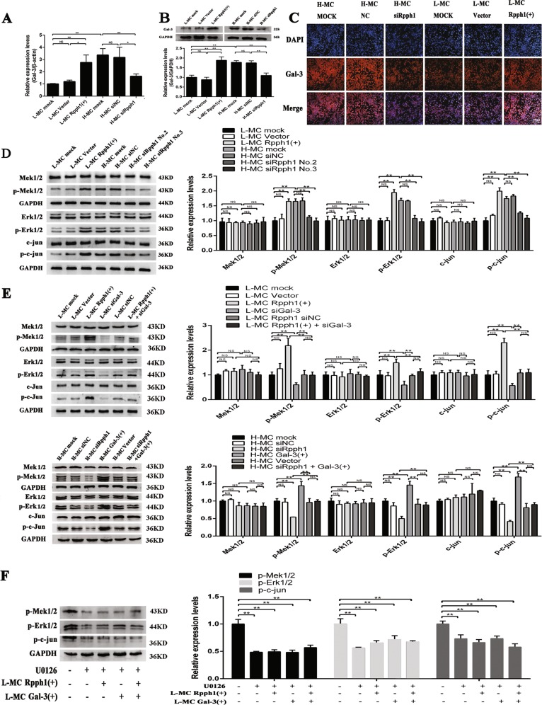
Fig. 5. Rpph1 (ribonuclease P RNA component H1) regulates activation of the Gal-3/Mek/Erk signaling pathway in mesangial cells (MCs).
a Twenty-four hours after transfecting Rpph1 small interfering RNA (siRNA) in H-MC or over-expression plasmid in L-MC, the messenger RNA (mRNA) levels of Gal-3 were detected by quantitative real-time PCR (qRT-PCR). b Forty-eight hours after transfecting Rpph1 siRNA in H-MC or over-expression plasmid in L-MC, Western blot and quantitative analysis were performed to analyze the protein levels of Gal-3. c Forty-eight hours after transfecting Rpph1 siRNA in H-MC or over-expression plasmid in L-MC, immunofluorescence was performed to analyze the protein level of Gal-3. d Forty-eight hours after transfecting Rpph1 siRNA in H-MC or over-expression plasmid in L-MC, Western blot and quantitative analysis were performed to analyze the expression of the key factors of the Mek/Erk signaling pathway. The antibodies of Mek, Erk, and c-Jun were stripped after the development by the antibody stripping solution. Then, the antibodies of p-Mek, p-Erk, and p-c-Jun were incubated with these bands for re-blotting, respectively. e The rescue experiment was performed to detect the effect of Rpph1 on activation or inhibition of the Mek/Erk signaling pathway. Up: Forty-eight hours after transfection with Rpph1 over-expression plasmid or co-transfection of Rpph1 over-expression plasmid and Gal-3 siRNA in L-MC, the expression of key factors of the Mek/Erk pathway were detected by Western blot and quantitative analysis. Low: Forty-eight hours after transfection with Rpph1 siRNA or co-transfection of Rpph1 siRNA and Gal-3 over-expression plasmid in H-MC, the expression of key factors of the Mek/Erk pathway were detected by Western blot and quantitative analysis. The antibodies of Mek, Erk, and c-Jun were stripped after the development by the antibody stripping solution. Then, the antibodies of p-Mek, p-Erk, and p-c-Jun were incubated with these bands for re-blotting, respectively. f Twenty-four hours after treatment with U0126 (10 μM) in L-MC, Western blot, and quantitative analysis were performed to analyze the effect of Rpph1 on regulation of the activation of Mek/Erk pathway; regulation was interrupted by U0126.The antibody p-Mek and glyceraldehyde-3-phosphate dehydrogenase (GAPDH) was stripped after the development by antibody stripping solution and then the antibodies of p-Erk and p-c-Jun was incubated with the bands, respectively. In all panels, the data are representative of three independent experiments. Data are presented as mean ± SD. *P < 0.05, **P < 0.01, NS not significant

