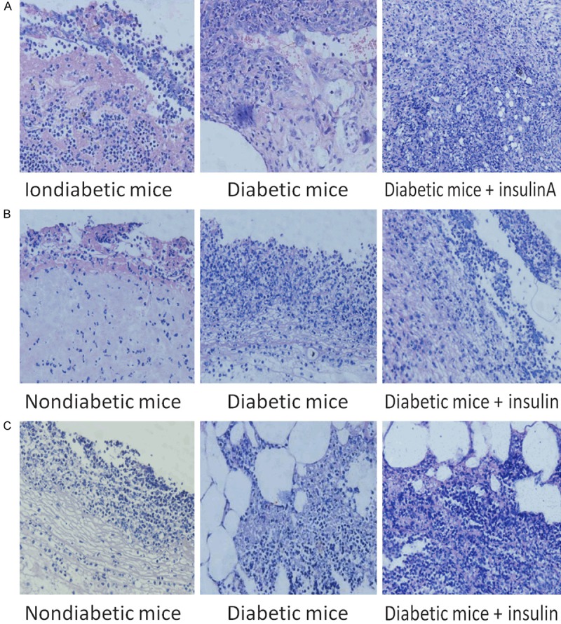
Figure 6.

Deparaffinized pathological tissue samples stained with HE to observe the inflammatory reactions of the cells in the infected sections. Arrows indicate P. aeruginosa cells observed by microscopy. Tissues infected with the (A) PAO1 wild-type strain, (B) PAO1/Plac-yhjH strain and (C) PAO1ΔwspF strain.
