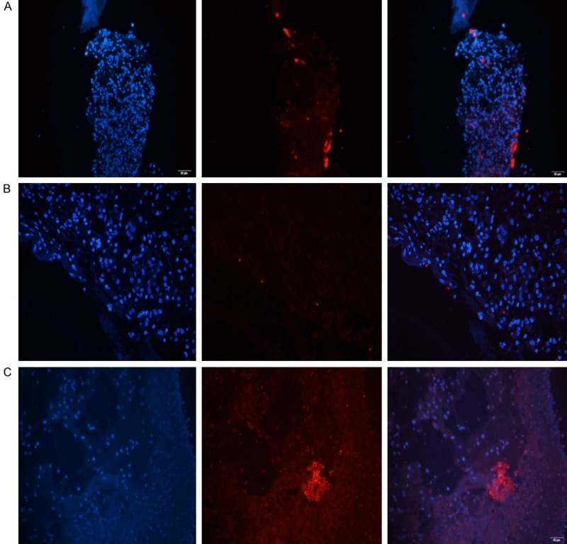
Figure 7.

P. aeruginosa aggregates were observed by PNA-FISH with a Texas Red-labeled P. aeruginosa-specific probe (red). The (A) PAO1 wild-type strain, (B) PAO1/Plac-yhjH strain and (C) PAO1ΔwspF strain observed with a fluorescence microscope. A high level of P. aeruginosa aggregation was observed with the PAO1ΔwspF strain (with high intracellular levels of c-di-GMP). Some scattered P. aeruginosa aggregates were observed with the PAO1/Plac-yhjH strain (with low intracellular levels of c-di-GMP).
