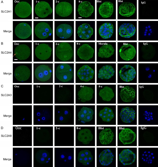
Figure 3.
Expression of SLC2A in the oocytes and preimplantation embryos from the reproductive tract of mouse analyzed by confocal microscopy. The images of SLC2A1, SLC2A3, SLC2A4, and SLC2A8 obtained by Hoechst 33342 staining of DNA are representative of 20 oocytes or embryos at different stages. Non-immune IgG equivalent was used as a negative control for the primary antibody (four-cell embryos and blastocysts as examples). Scale bar = 10 µm for all images.

