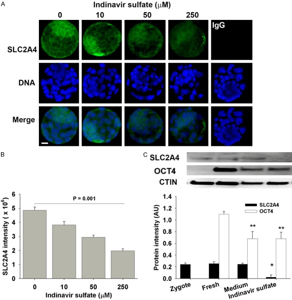
Figure 5.
The effect of indinavir sulfate on SLC2A4 in blastocyst. The whole-session epi-immunofluorescent images of SLC2A4 staining (A) and intensity (B) in the blastocysts derived from hybrid zygotes in KSMO medium supplemented with or without indinavir sulfate. The results were representative of three independent replicates with at least 30 embryos in each treatment. Negative control was the staining of non-immune IgG in blastocyst (medium only as example). Scalar bar = 10 for all images. (C) Western blot analyzed the expression of SLC2A4 and OCT4 in zygotes, fresh blastocyst (collected from reproductive tracts), and the blastocysts derived in KSOM medium or supplemented with 250 µM indinavir sulfate from zygote stage. The results were representative of three independent replicates with 70 embryos loaded in each lane. *P = 0.001, compared to fresh or medium-derived blastocysts. **P < 0.01, compared to fresh blastocysts.

