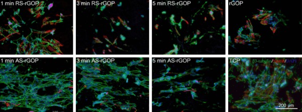
Figure 3.
Immunostaining images of SH-SY5Y cells growing on the arranged directional matrix of nanofibers with staining for DAPI (blue), β3-tubulin (green), and Nestin (red). The upper row of images shows the differentiated cells that were supported on randomly oriented silk nanofiber-reduced graphene oxide paper (RS-rGOP) after electrospinning for 1, 3, and 5 min, with rGOP as the control. The lower row of images shows the differentiated cells growing on AS-rGOP after different electrospinning for 1, 3, and 5 min, with tissue culture plate as the control. The neuron-specific marker β3-tubulin was expressed to the greatest extent on AS-rGOP with an electrospinning time of 1 min.

