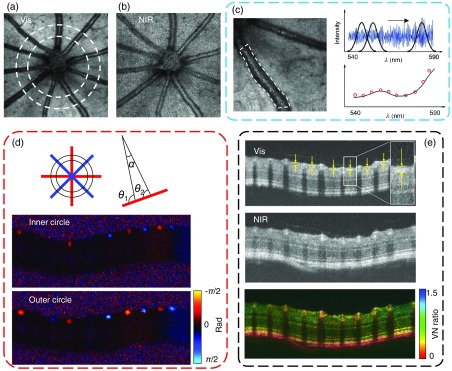
Fig. 2.
Multimetric measurements by vnOCT. (a), (b) En face projection of mouse retina in the visible and NIR channels, respectively. Two concentric circles represent the dual-circle scanning pattern. (c) Method for calculation by vessel segmentation and subsequent spectroscopic analysis. The vessel location was first manually segmented, and short-time Fourier transform was performed to obtain 4-D data (). Finally, the spectra from the bottom vessel wall were averaged and fitted by Eq. (1). (d) The schematic of blood flow measurements. The difference of the phase contrast from the inner and outer circles was used to calculate Doppler angle and blood velocity. (e) Illustration of RNFL thickness and RNFL-RRC VN ratio calculation. The circular B-scan image in visible light OCT was used to calculate RNFL thickness, as shown in yellow arrows. The intensity ratio between the visible and NIR channels was defined as VN ratio.

