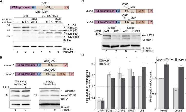
Figure 3.
A premature termination codon at position Q52′ enables the expression of Δ66′p53 and stabilises the mRNA transcript. (A) The effect of initiation from the AUG codons at positions 40′ and 44′ on the level of reinitiation from the AUG codon at position 66′. Western blot analysis of total cell lysates of HEK293T cells expressing the indicated variants of WT p53 (lanes 1–3) or Q52′TAG p53 (lanes 4–6). (B) The effect of intron 5 on the expression of Δ66′p53. Expression of N-truncated p53 variants in HEK293T cells, transiently (left) or stably (right) transfected with p53 Q52′TAG cDNA (–intron 5, lane 1) or Q52′TAG minigene (+intron 5, lane 2), were evaluated by Western blot analysis using antibodies against the C-terminal HA tag. (C) Knockdown of hUPF1. HEK293T cells stably expressing Q52′TAG minigene with either Met or Leu at position 66′ were treated with siRNA directed against hUPF1 or control siRNA (cont.). Protein expression levels were evaluated by immunoblotting using a specific antibody against hUPF1. (D) Left: Levels of SC35 1.7, CARS, SMG1 and p53 mRNA transcripts measured by RT-qPCR, following transfection of cells with sequence-specific or control siRNA, as described in panel C. Data represent the fold change in mRNA levels of the indicated transcript, relative to control. Right: Relative levels of mRNA transcripts measured in cells stably expressing the indicated Q52′TAG minigene and treated with hUPF1-specific or control siRNA. Data are displayed relative to mRNA levels of Q52′TAG minigene in cells treated with control siRNA. Values represent the mean ± SD of at least three independent experiments, each measured in triplicates.

