Figure 2.
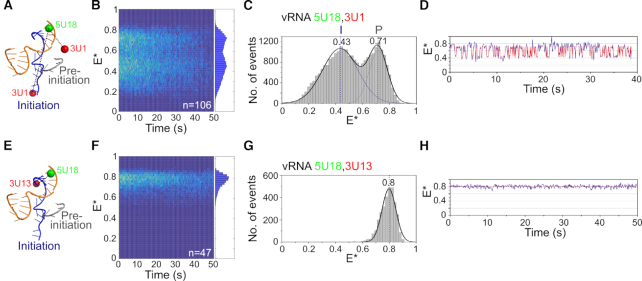
The 3′ vRNA in immobilized replication complexes is dynamic. (A) Schematic of conformations of the vRNA promoter in the absence of nucleotides, labeled with donor and acceptor fluorophores at positions 18 on the 5′ end and 1 on the 3′ end. (B) Kymograph of single-molecule time traces from immobilized replication complexes in the absence of nucleotides. E* represents apparent FRET efficiency. (C) FRET histogram from (B) fitted with a double Gaussian function. Initiation state = I, pre-initiation state = P. Frame time: 100 ms. (D) Example time trace showing transitions between the initiation and pre-initiation states. (E) Schematic of the vRNA promoter labeled with donor and acceptor fluorophores at positions 18 on the 5′ end and 13 on the 3′ end. (F) Kymograph of single-molecule time traces from immobilized replication complexes in the absence of nucleotides. (G) FRET histogram from (F). (H) Example time trace showing static FRET.

