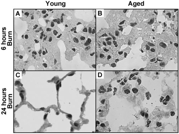
Fig. 2.
High-power view of H&E lung sections illustrating neutrophils within thickened alveolar walls of young and aged, burn-injured mice at 6 h (A andB) and at 24 h (C and D). High-power images of young, burn-injured mice at 24 h did not appear different from those of sham-injured mice (not shown). All images are at 1000 × original magnification.

