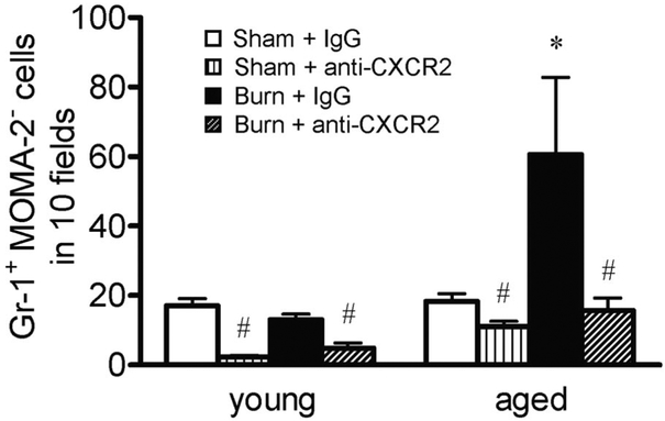
Fig. 7.
Total numbers of Gr-1+ MOMA-2− cells from lungs of young and aged animals at 24 h after sham or burn injury, receiving 20 μg i.p. control IgG or 20 μg i.p. anti-CXCR2 antibody, were counted in sections of lung tissue as described above. Data are represented as the average number of cells counted in 10 400x fields for each group ± SEM. The average tissue area over which cells were counted did not differ between groups; n =3–9 mice per group; *, P < 0.05, compared with all other groups; #, P < 0.05, compared with IgG controls.

