FIGURE 5:
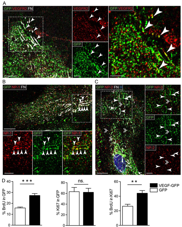
VEGFR2 and NP-1 but not NP-2 shows clustering and colocalization with VEGF-GFP at cell-matrix adhesions. Representative confocal images of astrocytes expressing VEGF-GFP immunostained for GFP, FN and (A) VEGF receptor 2 (VEGFR2), or its coreceptors (B) neuropilin-1 (NP-1), and (C) neuropilin-2 (NP-2). All three proteins had a diffuse, dot-like distribution in the cells, and additionally, VEGFR2 and NP-1 formed linear clusters at FN+ adhesions (arrowheads in A and B), while NP-2 did not (arrowheads in C). Colocalization between VEGF-GFP and VEGFR2 or NP-1 was restricted mainly to linear clusters at cell-matrix adhesions (arrowheads in A and B), while NP-2 occasionally colocalized with VEGF-GFP in vesicle-like structures (double arrowhead in C). H, Hoechst; Bars: 10 μm (main) and 5 μm (close up). D: Quantifications following BrdU pulse labeling of subconfluent cultures in serum free medium demonstrated more BrdU+ cells among astrocytes expressing VEGF-GFP than in control GFP cells (% BrdU in GFP). The proliferative fraction (% Ki67 in GFP) was equal at both conditions, meaning that the cell cycle was accelerated by VEGF-GFP (% BrdU in Ki67). This mitogenic effect suggests the presence of VEGFR2 signaling initiated by VEGF-GFP in astrocytes. n = 6, unpaired t test, ** P<0.01, *** P<0.001 (see also Supp. Info. Fig. 3)

