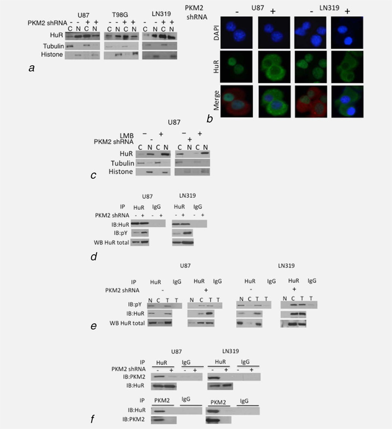
Figure 3.

PKM2 controls the sub-cellular localization of HuR. (a, b) Levels of HuR, histone H3 and tubulin in the cytoplasmic (C) and nuclear (N) fractions of cells expressing a scramble or PKM2-targeted shRNA as assessed by Western blot (a) and DAPI(blue)/HuR (green) co-immunofluorescence analysis (b). PKM2 staining (red) delineates the cytoplasm in groups with minimal cytoplasmic HuR. (c) Western blot analysis of HuR, tubulin and histone in nuclear and cytoplasmic fractions of control and shPKM2 expressing U87 cells incubated with 0 or 5ng leptomycin B (LMB, 8 hr). (d) Western blot analysis of HuR and pY proteins in total cellular HuR or IgG immunoprecipitates from control or PKM2-suppressed U87 and LN319 cells. (e) Western blot analysis of HuR and pY proteins in total cellular (T) nuclear (N), or cytoplasmic (C) HuR or IgG immunoprecipitates from control or PKM2-suppressed U87 and LN319 cells. (f) Western blot analysis of PKM2 and HuR levels in HuR immunoprecipitates, and HuR levels in PKM2 immunoprecipitates from U87 and LN319 cells expressing a scramble or PKM2-targeted shRNA.
