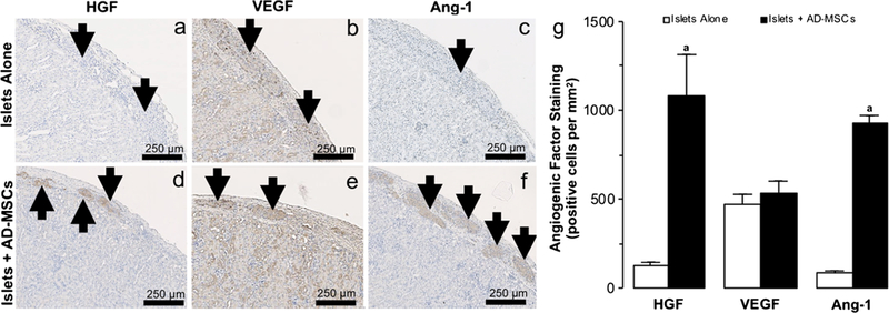
Fig. 5.

Angiogenic factors within transplanted islets. a–f Representative images following hepatocyte growth factor: HGF (a, d), vascular endothelial growth factor: VEGF (b, e), and angiopoetin-1: Ang-1 (c, f) immunohistochemical staining of 150 islets that were transplanted under the kidney capsule, either alone or with AD-MSCs, at 2 weeks following transplantation. Arrows indicate islets present within the representative images. g Quantification of the positive HGF, VEGF, and Ang-1 staining in samples where 150 islets were either transplanted alone (white square) or with AD-MSCs (black square). Significant differences: aP < 0.05 for islets alone vs. islets + AD-MSCs (Student’s unpaired t test)
