Fig. 5.
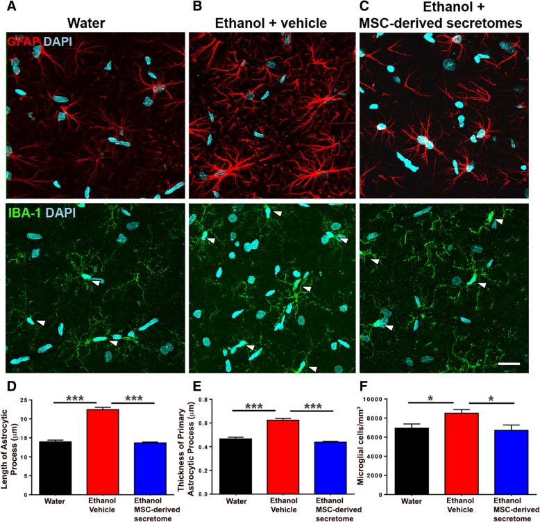
Intranasal administration of adipose tissue-derived MSC secretome normalized both the increased astrocyte reactivity and the increased microglial density induced by chronic ethanol intake. Representative confocal microphotographs of hippocampal astrocyte GFAP immunoreactivity (red, top) and microglial density (Iba-1; green, shown by arrows, center) immunoreactivity counterstained with DAPI (blue, nuclear marker) (scale bar 25 μm). Chronic ethanol-drinking rats treated with vehicle displayed a marked increase in the length and thickness of astrocyte processes (b, top) and microglial density (b, center) versus rats drinking only water (a, top and center). Administration of five intranasal doses of secretome, administered at weekly intervals (blue bar), to chronic ethanol drinking rats normalized the length and thickness of astrocytic process and microglial density (c, top and center). One-way ANOVA of ethanol-drinking vehicle-treated rats versus water-drinking rats respect to (i) the length of astrocyte processes (d); F2, 1984 = 153.6, p < 0.0001; Tukey post-hoc: p < 0.001; (ii) the thickness of primary processes (e); F2, 727 = 93.04, p < 0.0001; Tukey post-hoc: p < 0.001; and (iii) microglial cell density/mm3 (f); F2, 56 = 5.246, p < 0.05; Tukey post-hoc: p < 0.001. One-way ANOVA ethanol-drinking rats secretome-treated versus ethanol-drinking rats vehicle treated respect to (i) the length of astrocyte processes (d); F2, 1984 = 153.6, p < 0.001; Tukey post-hoc: p < 0.001; (ii) the thickness of primary process (e); F2, 727 = 93.04, p < 0.001; Tukey post-hoc: p < 0.001; and (iii) microglial cell density/mm3 (f); F2,56 = 5.246; p < 0.01; Tukey post-hoc: p < 0.05

