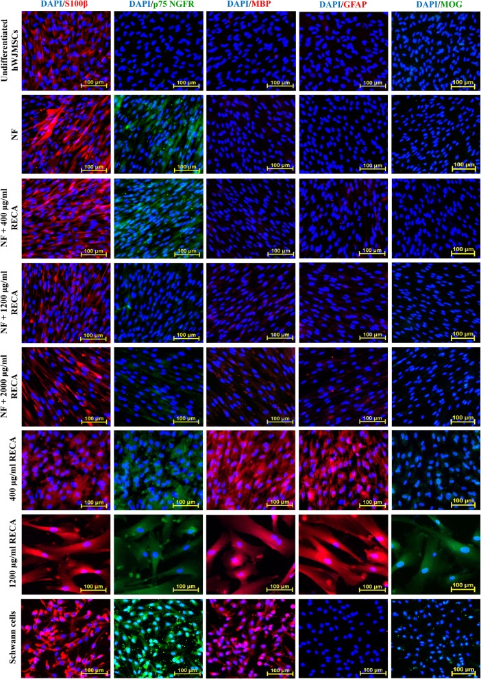
Fig. 9.
Immunocytochemical analysis of S100β, MBP, GFAP (red) as well as p75 NGFR and MOG (green) in differentiated and undifferentiated hWJMSCs. Cell nuclei were counterstained with DAPI (blue). Undifferentiated hWJMSCs served as a negative control, while Schwann cells acted as a positive control. hWJMSCs differentiated with 1200 μg/ml RECA showed prominent expression of all neural-specific markers compared to differentiated hWJMSCs in other induction groups (n = 6). DAPI; 4′, 6-diamidino-2-phenylindole. Scale bar 100 μm

