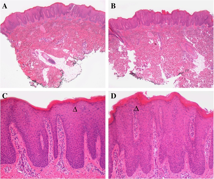
Fig. 2.
Microscopic evaluation of twin skin biopsies. At different magnification, microscopic evaluation of Haematoxylin&Eosin-stained paraffin sections of skin biopsies of both twins (a-c and b-d, respectively) reveals a parakeratotic cornified layer and epidermis with marked elongation of rete ridges and an almost absent granular layer (arrow heads); in the papillary dermis, inflammatory cells surround dilated tortuous small vessels, consistent with the diagnosis of psoriasis. Haematoxylin&Eosin stain; original magnification a, b: 40X; c, d: 100X

