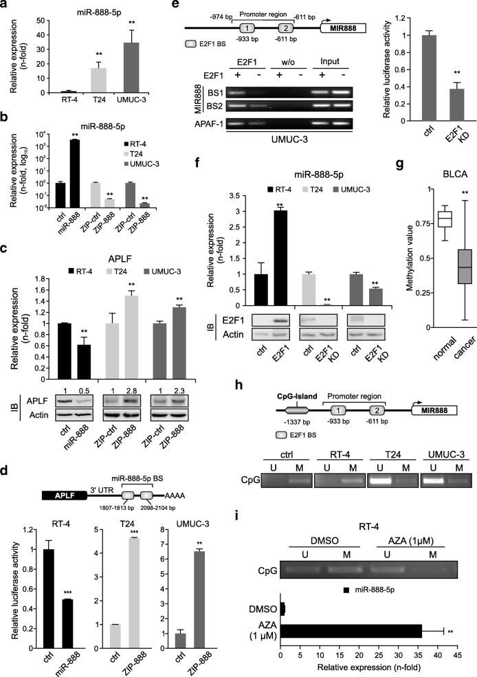
Fig. 4.
E2F1 accesses the hypomethylated MIR888 and transactivates the APLF inhibitor miR-888-5p in invasive BC. a Validation of endogenous miR-888-5p transcript levels in RT-4, T24 and UMUC-3 cells. MiRNA expression in RT-4 cells was set as 1. b, c qPCR of miR-888-5p (b) and APLF levels (c, top) in RT-4 expressing mir-888-5p (black), in T24.ZIP-888 (light grey) and in UMUC-3.ZIP-888 (dark grey) versus controls. Immunoblots (IB) of APLF are shown below with actin as loading control. d Scheme of the APLF 3’UTR with predicted miR-888-5p binding sites (BS). Reporter assays with above-mentioned cell lines co-transfected with the 3’UTR luciferase construct. Controls were set as 1. e Top: Scheme of the MIR888 promoter region with two predicted E2F1 binding sites. Bottom: ChIP assays for MIR888 promoter in E2F1-depleted UMUC-3 cells using anti-E2F1 antibody. Mock IPs (w/o) of the samples were used as a negative control, while input was used as positive control. The APAF-1 promoter was used as an E2F1 binding control. Right: The promoter activity was measured using a luciferase assay and the control was set as 1. f Quantification of miR-888-5p in indicated BC cell lines after overexpression or knockdown (KD) of E2F1. E2F1 protein levels were validated using immunoblots (bottom), using actin as loading control. g Methylation status of MIR888 promoter region ranging 2000 bps upstream of TSS in Bladder Urothelial Carcinoma (BLCA). h Top: MIR888 promoter region with the CpG island in proximity to the E2F1 binding sites. Bottom: Methylation-specific PCR in RT-4, T24, UMUC-3 cells using primers that discriminate between the unmethylated (U) and methylated (M) CpG island upstream of MIR888 TSS. The human methylated DNA standard (ctrl) was used as positive control. i Methylation-specific PCR for MIR888 CpG island in AZA-treated RT-4 cells (top) and validation of miR-888-5p levels (bottom). Asterisks indicate statistically significant (** p < 0.01, *** p < 0.01) changes

