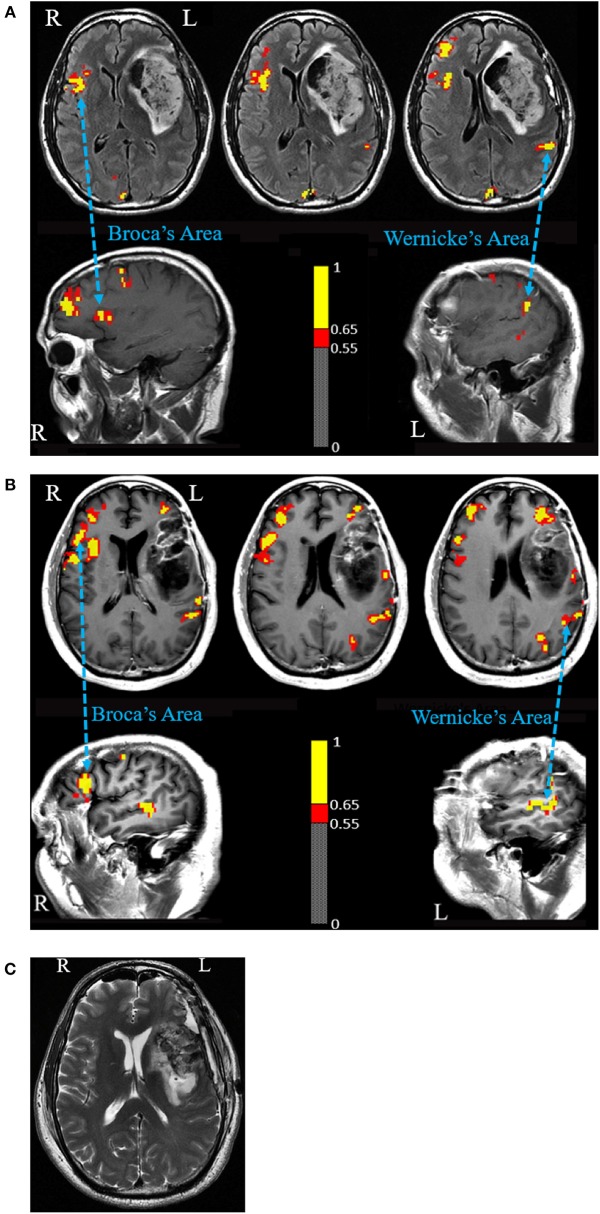
Figure 1.
(A) Language fMRI prior to first surgery. A semantic fluency task fMRI shows activated Broca's Area homolog in the right hemisphere and Wernicke's Area in the left hemisphere. The figure shows voxels activated at a correlation coefficient of 0.54 (p = 3.8 × 10⁻⁸) or higher. (B) Language fMRI prior to second surgery. A verbal fluency task shows activated Broca's Area homolog in the right hemisphere and Wernicke's Area in the left hemisphere. Activation was also seen anterior and posterior to the expected Broca's Area in the left hemisphere. Intraoperative cortical stimulation elicited no speech disturbance in the anterior margin and motor-related disturbances in the posterior margin. The figure shows voxels activated at a correlation coefficient of 0.55 (p = 1.7 × 10⁻⁷) or higher. (C) Post-operative MRI scan shows a gross total resection including the expected anatomical region of Broca's Area.

