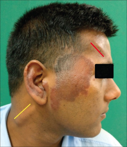
Figure 1.

Malar region (Rt) and ear showing well-defined erythematous edematous plaque, Grade 3 thickened (Rt) supraorbital (red line), and greater auricular nerve (yellow line)

Malar region (Rt) and ear showing well-defined erythematous edematous plaque, Grade 3 thickened (Rt) supraorbital (red line), and greater auricular nerve (yellow line)