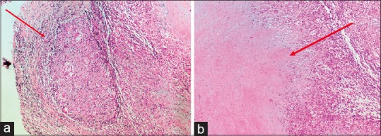
Figure 3.

(a) Nerve biopsy of greater auricular nerve (Rt) showing intact nerve within perineurium with multiple ill-formed granulomas bordered by epithelioid cells, lymphocytes, and giant cell reaction (10×, H and E stain). (b) Nerve biopsy revealing large areas of caseous necrosis surrounded by granuloma characteristic of segmental necrotizing granulomatous neuritis (10×, H and E stain)
