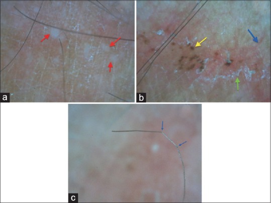
Figure 3.

(a) Micropustules (red arrow) seen over a background of erythema and scaling; (b) dermoscopy of a ring of T. pseudoimbricata showing scaling (green arrow), linear vessel (blue arrow), and brown hemorrhagic spot (yellow arrow); (c) vellus hair in the lesion appear bent and broken due to fungal invasion of the hair shaft [Dinolite AM413ZT; 200X; Polarising]
