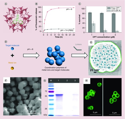
Figure 2. . In situ encapsulating drug molecules into nanoscale metal-organic frameworks.
(A) Topological structure of the fluorescein-loaded ZIF-8 framework. (B) pH-responsive fluorescein release profiles in PBS solution. (C) Cell viability of MCF-7 cells incubated with free CPT and CPT loaded ZIF-8 nanoparticles. (D) pH-induced in situ synthesis of drug-loaded zeolitic imidazolate framework-8 (ZIF-8) nanoparticles. (E) Cross-section of the electron tomogram of mesoporous structure distribution in the doxorubicin-loaded ZIF-8 nanoparticles. (F) Scanning electron microscope image of catalase-loaded ZIF-90 nanoparticles. (G) SDS-PAGE gel image (M: protein marker, lane 1: free catalase, lane 2: washed ZIF-90 with catalase on the surface, lane 3: catalase-loaded ZIF-90). (H) Confocal microscopy images of fluorescently (FITC)-labeled catalase-loaded ZIF-90 sample (left) and ZIF-90 with fluorescein isothiocyanate-catalase on its surface (right).
CPT: Camptothecin; ZIF-8: Zeolitic imidazolate framework-8.
(A) Reproduced with permission from [42] © American Chemical Society (2014).
(B) Reproduced with permission from [42] © American Chemical Society (2014).
(C) Reproduced with permission from [42] © American Chemical Society (2014).
(D) Reproduced with permission from [47] © American Chemical Society (2016).
(E) Reproduced with permission from [47] © American Chemical Society (2016).
(F) Reproduced with permission from [44] © American Chemical Society (2015).
(G) Reproduced with permission from [44] © American Chemical Society (2015).
(H) Reproduced with permission from [44] © American Chemical Society (2015).

