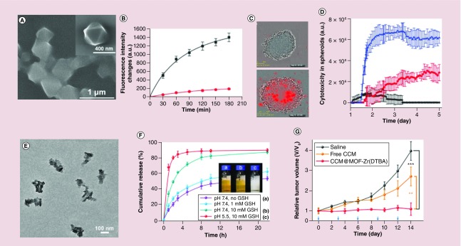
Figure 4. . Functional metal-organic framework nanocompositions for stimuli-responsive drug release.
(A) Scanning electron microscope image of a stimuli-responsive DNA-based polyacrylamide hydrogel-coated Universitetet i Oslo (UiO)-68 nanoparticles. (B) Release profiles of doxorubicin (DOX) from the hydrogel-coated UiO-68 nanoparticles upon addition of ATP (50 × 10-3 M, black line) and in absence of the ATP (red line). (C) Representative phase-contrast and red fluorescence images (indicative of the cell apoptosis) of the cell aggregates after time intervals of 24 h (top) and 120 h (down), where the cells were treated with DOX-loaded hydrogel-coated UiO-68 nanoparticles. (D) Apoptosis of the MDA-MB-231 cells treated with unloaded hydrogel-coated UiO-68 nanoparticles (black line), DOX-loaded UiO-68 nanoparticles gated by ATP-responsive duplex units (red line), and DOX-loaded ATP-responsive hydrogel-coated UiO-68 nanoparticles (blue line). (E) Transmission electron microscopy image of CCM@MOF-Zr (4,4′-DTBA) nanoparticles. (F) Glutathione-responsive release profiles from CCM@MOF-Zr (4,4′-Dithiobisbenzoic acid) nanoparticles in pH 5.5 and pH 7.4 phosphate-buffered saline solution. (G) Relative tumor volume and body weight of the mice.
CCM: Curcumin; DTBA: Dithiobisbenzoic acid; MOF: Metal-organic framework.
(A) Reproduced with permission from [75] © WILEY‐VCH Verlag GmbH & Co. KGaA, Weinheim (2016).
(B) Reproduced with permission from [75] © WILEY‐VCH Verlag GmbH & Co. KGaA, Weinheim (2016).
(C) Reproduced with permission from [75] © WILEY‐VCH Verlag GmbH & Co. KGaA, Weinheim (2016).
(D) Reproduced with permission from [75] © WILEY‐VCH Verlag GmbH & Co. KGaA, Weinheim (2016).
(E) Reproduced with permission from [76] © American Chemical Society (2018).
(F) Reproduced with permission from [76] © American Chemical Society (2018).
(G) Reproduced with permission from [76] © American Chemical Society (2018).

