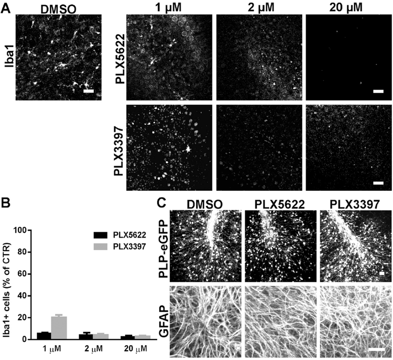
Fig.1.

CSF1R inhibitors PLX5622 and PLX3397 effectively depleted microglia without affecting oligodendrocytes or astrocytes in cerebellar slices. PLX5622 or PLX3397 were applied to cerebellar slices prepared from PLP-eGFP pups at various concentrations as indicated for 3 days. A. Representative confocal images of Iba1 immunostaining in control (DMSO), PLX5622 or PLX3397 treated slices. B. Quantification of the number of Iba1+ cells in slices for all treatment groups. Cell count was normalized to DMSO control (CTR) slice. n=3-4. C. Confocal images of PLP-eGFP and GFAP immunostaining in slices treated with DMSO or 2 μM PLX5622 or PLX3397. Scale bars: 25 μm.
