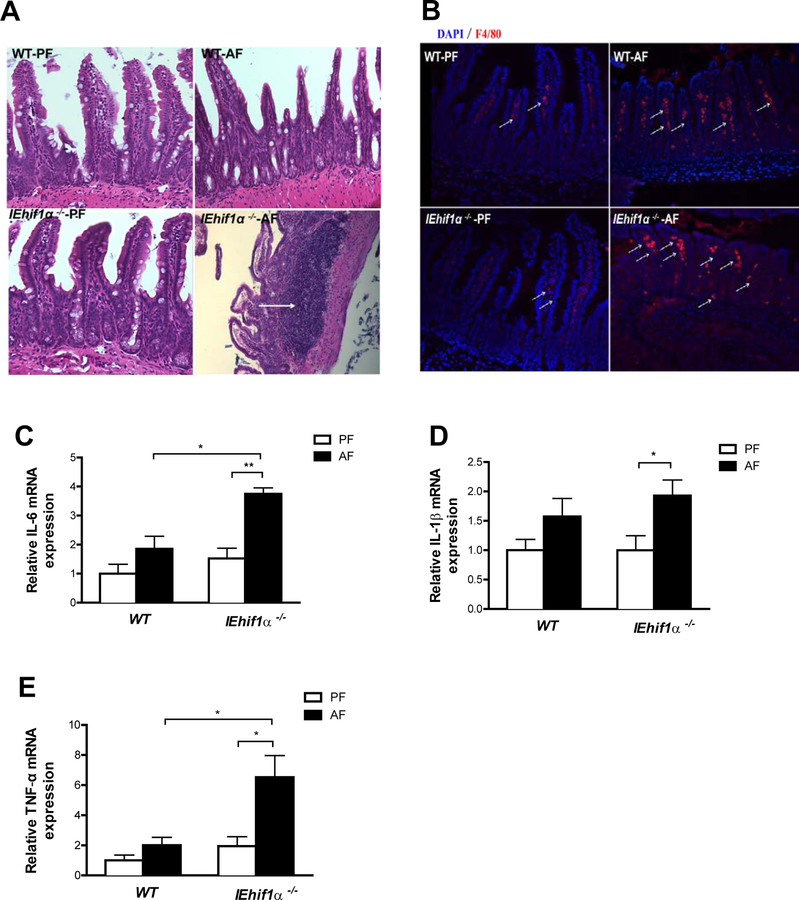
Figure 6.
Effect of Hif-1α on intestinal inflammation. Mice were treated as described in in Fig 2. (A) HE staining of ileal tissues. Original magnification, ×10. (B) Immunofluorescent staining of F4/80 (red) showed increased macrophages in ileum of AF IEhif1α−/− mice (arrows). DAPI (blue) was used to visualize nuclei. Original magnification: ×10. (C, D, E) Ileum IL-6, IL-1β and TNF-α mRNA levels. Data are expressed as mean ± SEM. AF, alcohol-fed; PF, pair-fed.

