
Figure 2.
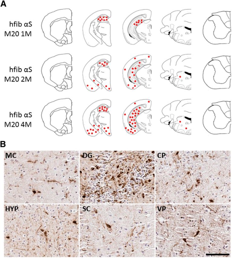
Induction of pSer129/81A-stained inclusions in M20 Tg mice after intrahippocampal injection of 21–140 hfib αS. A, Schematic map showing rostrocaudal distribution of inclusions detected with pSer129/81A antibody in M20 Tg mice at 1, 2, and 4 months after injection. Similar density and distribution of inclusion pathology was seen bilaterally. Inclusions were mainly localized around the site of injection (hippocampus) at 1 month after injection. At 2 and 4 months after injection, more abundant inclusion formation was observed in the hippocampus, and it had progressed to the cortex, striatum, midbrain, and brainstem. B, Representative images of immunohistochemistry showing regions with pSer129/81A-immunoreactive inclusions at 4 months after injection. Perinuclear, intracellular aggregates and neuritic pathology extending into processes are shown in the motor cortex (MC), hippocampus (dentate gyrus; DG), caudate–putamen (CP), hypothalamus (HYP), superior colliculus (SC), and ventral pons (VP). Tissue sections were counterstained with hematoxylin. Scale bar: MC, 100 μm; DG, CP, HYP, SC, and VP, 50 μm.