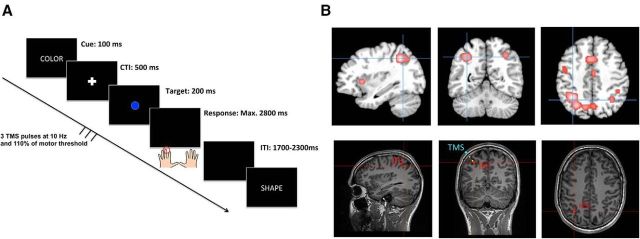
Figure 5.
Illustration of the setup of Experiment 3. Single-trial structure of the experimental paradigm (A). In this experiment, participants completed only TGBs using their left and right middle fingers throughout all blocks. The TMS pulses were time-locked to the onset of the target stimulus to affect the final preparation stages. To avoid stimulation at the time of a response, the number of pulses was reduced to three pulses. Localization of the parietal target site (B) was identical as to Experiment 2 (MNI coordinate: −34, −56, 43). The coronal sections of the bottom also display the position of the coil center, where the magnetic field is maximal, relative to the subject's skull (blue markers). Yellow spheres indicate the direction of the current flow (distance between two spheres = 1 cm). The average distance between the skull and the target site was 2.41 cm across participants. Note that the application of TMS trains also affects the brain tissue between the skull and the target site.

