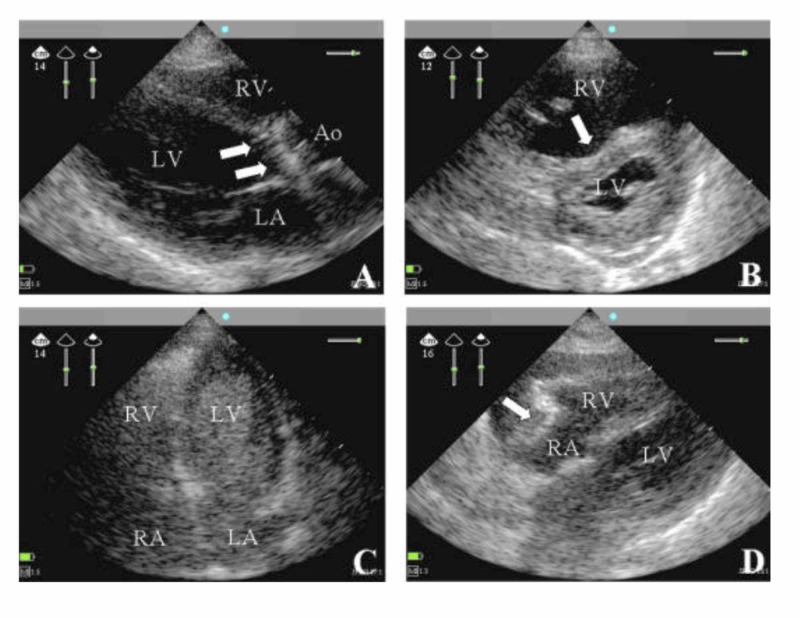
Figure 1. (Panels A-D) Views from Four Different Patients.
(Panel A) The parasternal long-axis (PLA) view was taken from a patient with septic shock. The patient had severe aortic endocarditis (arrows pointing to vegetations present on the aortic valve) and a dilated left ventricle. The aorta (Ao), the right ventricle (RV), and the left atrium (LA) are also seen [6].
(Panel B) The parasternal short-axis (SA) view was taken from a patient with acute respiratory distress syndrome and associated right heart dysfunction. The right ventricle (RV) is shown to be enlarged due to severe pulmonary hypertension (seen with arrow) [6].
(Panel C) The apical (AP) view was taken from a ventilated patient with refractory hypoxemia. The right ventricle (RV), the left ventricle (LV), the right atrium (RA), and the left atrium (LA) all are seen [6].
(Panel D) The subxiphoid (SX) view was taken from a patient with shock and pulsus paradoxus. Pericardial effusion is present (pointed out by the arrow). Also seen are the right ventricle (RV), the right atrium (RA), and the left ventricle (LV) [6].

