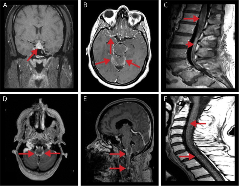
Figure 2. Examples of typical NS MRI findings.
(A) Coronal T1 postgadolinium pituitary and pituitary stalk lesion with enhancement (arrow). (B) Axial T1 postgadolinium leptomeningeal contrast enhancement primarily about the base of the brain (in a patient with probable NS, known to have biopsy-confirmed pulmonary sarcoidosis; arrows). (C) Midsagittal T1 postgadolinium of lumbar spine thickening and smooth/nodular leptomeningeal enhancing lesions surrounding the conus medullaris (arrow) and cauda equina (arrowhead). (D) Axial T1 postgadolinium leptomeningeal enhancement of the midbrain (arrows). (E) Sagittal postgadolinium nodular leptomeningeal enhancement of the lower brainstem (arrows). (F) Midsagittal T1 postgadolinium of cervical spine multiple nodular leptomeningeal enhancing lesions (arrows; in a patient with probable NS, known to have biopsy-confirmed pulmonary sarcoidosis). NS = neurosarcoidosis.

