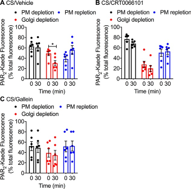
Figure 6.

CS-induced trafficking of PAR2-Kaede from the Golgi apparatus to the plasma membrane. PAR2-Kaede located in the region of the perinuclear Golgi apparatus of KNRK-PAR2-Kaede cells was green/red photoconverted. Cells were then incubated with CS (100 nm) for 0 or 30 min. PAR2-Kaede fluorescence was measured at the cell surface or Golgi. Cells were pre-incubated with vehicle (A), PKD inhibitor (CRT0066101, 100 nm) (B), or Gβγ inhibitor (gallein, 10 μm) (C). Quantification of PAR2-Kaede (518 nm, green) at the plasma membrane and of photoconverted PAR2-Kaede (572 nm, red) in the Golgi region and at the plasma membrane is shown. n = 6 experiments, >30 cells analyzed per condition. *, p < 0.05 (Student's t test). Error bars, S.E.
