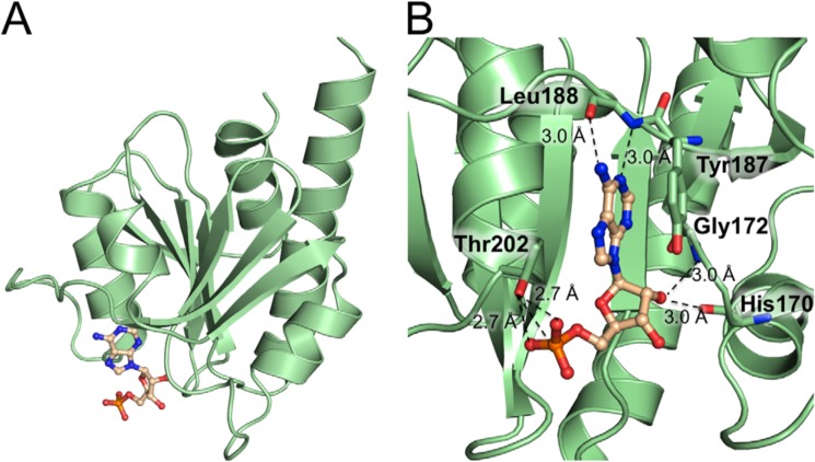
Figure 4.
Structure of the CdaA-AMP complex. A, CdaA monomer (cartoon, pale green) with a bound AMP molecule depicted as balls and sticks. B, a detailed view of the active site showing the amino acids (sticks, carbon in pale green, oxygen in red, and nitrogen in blue) involved in AMP binding. The bound AMP is shown as a ball-and-stick model (carbon in wheat, phosphate in orange, oxygen in red, and nitrogen in dark blue).

