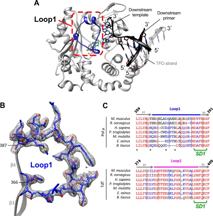
Figure 5.
Ordered structure of Loop1 in TdT-μ chimera bound to the downstream DNA duplex and the incoming dNTP. A, overview of the ternary complex. Loop1 is shown in deep blue and boxed in a red dotted frame. The DNA duplex is in black. The incoming nucleotide is in green. B, electron density in the 2Fo − Fc map (contoured at 1σ in gray) in the Loop1 region of TdT-μ chimera pre-ternary complex. Loop1 is represented in a ball-and-stick model in the context of the adjacent β3 and β4 strands, shown in gray. C, sequence alignments of TdT and Pol μ in the Loop1 region. Loop1 of Pol μ and TdT are delimited with a light blue and pink line, respectively. The interactions between side-chain atoms of Loop1 and the remaining part of TdT are indicated with an asterisk. Strictly conserved residues are shown in red, and residues with similar physico-chemical character are in blue.

