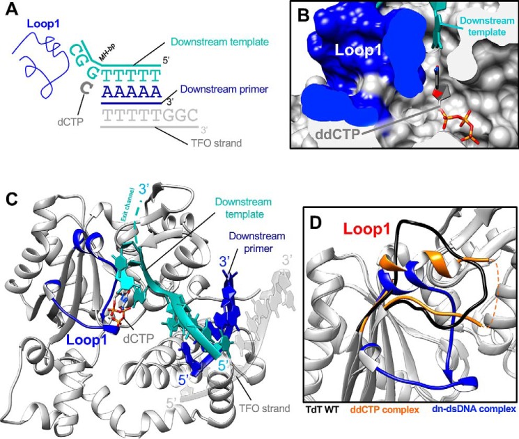
Figure 7.
Structure of TdT chimera as a ternary complex with a downstream dsDNA and the incoming dNTP. A, DNA substrates; the downstream DNA duplex is colored in blue and cyan. The incoming nucleotide and Loop1 are represented in dark gray and blue, respectively. The additional DNA strand, a triplex-forming oligonucleotide (TFO) is represented in gray. B, space-filling representation of the incoming ddNTP-binding site. The ddCTP is in ball-and-stick form, the downstream template strand is in cyan, and the surface of the Loop1 atoms is in dark blue. C, overall structure of the TdT-μ chimera downstream dsDNA. The incoming ddCTP makes Watson–Crick interactions with the in trans template strand. Loop1 is fully visible in the electron density map and is made of two 310 helices and one α helix. The exit channel for the 3′ protruding end is represented by a dashed cyan line. D, differences in the Loop1 conformation in the binary ddCTP complex (gold) and in the downstream dsDNA complex (dark blue). Loop1 conformation of TdT-WT apoenzyme is also represented as a reference (black).

