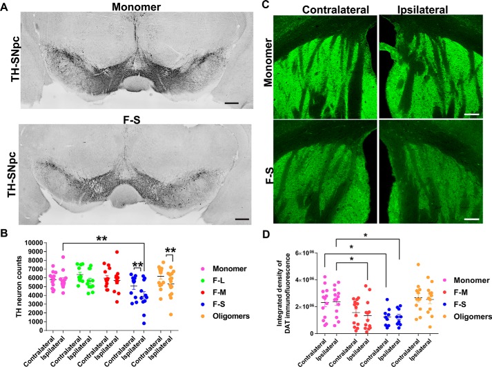
Figure 6.
Quantitation of TH-positive neurons in the SNc and DAT terminals in striatum following unilateral striatal injections of different α-synuclein species. C57BL/6J mice received unilateral striatal injections of 2 μl of soluble monomer (300 μm), F-L (150 μm), F-M (150 μm), F-S (150 μm), and O (300 μm). After 3 months, the mice were perfused, and immunostaining was performed. Numbers of mice are as follows: monomer (12); F-L (11); F-M (12); F-S (11); and O (15). A, representative images of tyrosine hydroxylase immunohistochemistry in the SNc of monomer- and F-S–injected mice. B, unbiased stereology of tyrosine hydroxylase–positive neurons performed by an investigator blinded to experimental conditions. Data are shown as the mean counts ± S.E. and analyzed using a two-way ANOVA, α-synuclein species F(5,67) = 2.7, p = 0.03. C, immunofluorescence performed using an antibody to DAT. Images were captured using confocal microscopy. Representative images of the striatum from monomer and F-S–injected mice are shown. D, ImageJ was used to quantify the integrated fluorescence intensity of DAT in the striatum. Data are shown as the mean counts ± S.E. and analyzed using a two-way ANOVA, α-synuclein species F(3,43) = 5.7, p = 0.002. *, p < 0.05; **, p < 0.01. Scale bar, 100 μm.

