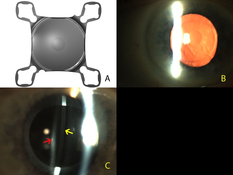
Figure 1.
(A) Digital high-resolution photograph of the Scharioth macula lens. Note the central zone with a +10.0 D addition. (B) Clinical photograph of the Scharioth macula lens implanted in the eye. Note the ‘oil droplet sign’ of the central +10.00 addition on the intraocular lens (IOL). (C) Slit lamp illustrating the second Purkinje reflection (yellow arrow) from the Scharioth macula lens in the ciliary sulcus and the third Purkinje reflection (red arrow) from the primary intraocular lens in the capsular bag. Note the dark band between the two reflections showing that there is a clear space between the two IOLs.

