
Figure 3.
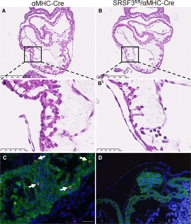
Loss of SRSF3 (serine/arginine splicing factor 3) in the developing heart results in embryonically lethal heart defects. A and B, Hematoxylin and eosin (H&E) staining in sections of αMHC-Cre embryos (A) and SRSF3-floxed/αMHC-Cre embryos (B) at E9.5. The boxed areas in A and B are shown at high magnification in A′ and B′, illustrating the cell deficit in the developing ventricular wall of SRSF3-floxed/αMHC-Cre embryos. Bar, 250 µm (A and B), 50 μm (A′ and B′). C and D, Immunofluorescence analysis in sections of αMHC-Cre embryos (C) and SRSF3-floxed/αMHC-Cre embryos (D) at E10.5, using anti-phospho-Histone H3 (red), anti-TroponinT (green), and Dapi (blue). Arrows indicate cardiomyocytes positive for phospho-histone H3; the developing SRSF3-floxed/αMHC-Cre heart is devoid of these cells. Bar, 20 μm.