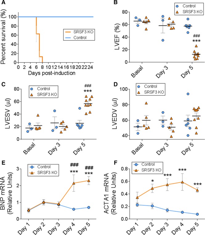
Figure 4.

Cardiac-specific SRSF3 (serine/arginine splicing factor 3) knockout (KO) mice develop severe and fatal contraction defects. Deletion of SRSF3 in cardiomyocytes was induced in inducible SRSF3 KO mice by 3 hydroxytamoxifen injections on alternate days (post-induction days 0, 2, and 4). A, Survival curves for hydroxytamoxifen-injected control and SRSF3 KO mice. B–D, Echocardiography analysis of left ventricular ejection fraction (B), left ventricular end-systolic volume (C), and left ventricular end-diastolic volume (D); ***P<0.001 SRSF3 KO vs Control, ###P<0.001 vs Basal; 2-way ANOVA followed by the Bonferroni post-test. E and F, qRT-PCR (polymerase chain reaction) analysis of the cardiac expression of BNP (E) and Acta1 (F) mRNA over 5 days post-induction. Data are shown as mean±SEM. n=5–13 mice per group. Two-way ANOVA followed by Bonferroni post-test. *P<0.05, ***P<0.001 SRSF3 KO vs Control. ###P<0.001 vs day 1. ACTA1 indicates actin alpha 1; BNP, brain natriuretic peptide; LVEDV, left ventricular end diastolic volume; LVEF, left ventricular ejection fraction; and LVESV, left ventricular end systolic volume.
