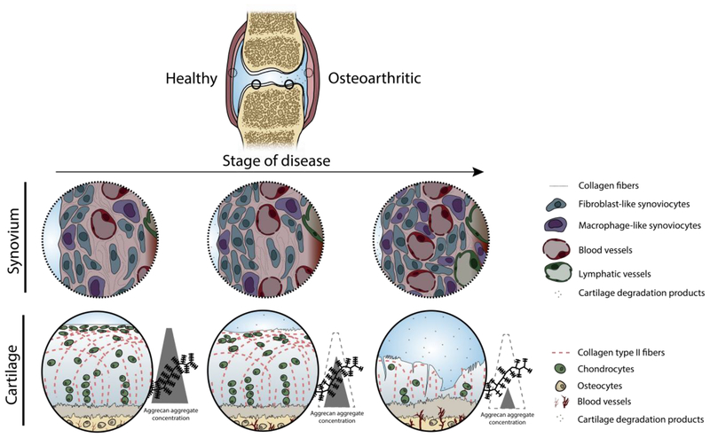
Figure 1.
Structural and compositional changes to the joint during OA disease progression, highlighting the changes to the cartilage and synovium. Synovium undergoes marked immune cell infiltration, increased vascularization, and increased vascular fenestration. Cartilage breakdown begins at the articular surface with proteoglycan fragmentation and loss, surface fibrillation, and chondrocyte disorganization and clustering at the articular surface [58]. In late stages of disease, the cartilage has lost bulk tissue as proteoglycans are depleted, the collagen network degraded by MMPs, deep fibrillation and fissures have developed, and the remaining chondrocytes are unable to repair at a sufficient rate.

