Figure 2.
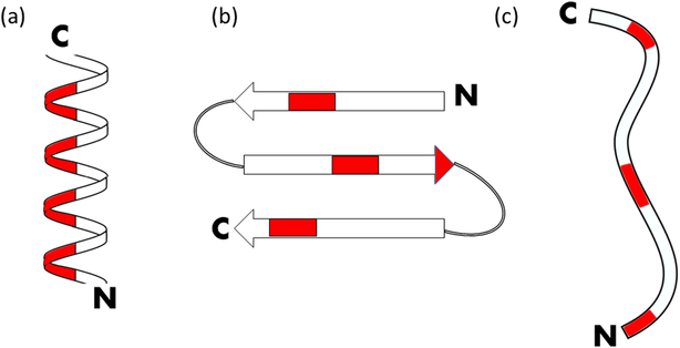
Antimicrobial peptides can be classified by their secondary structure: (a) α-helical, (b) β-sheet, (c) extended/flexible. Red regions indicate hydrophobic residues for the configuration of magainin, defensin 5, and indolicidin, respectively. N refers to the N-terminus, while C refers to the C-terminus.

