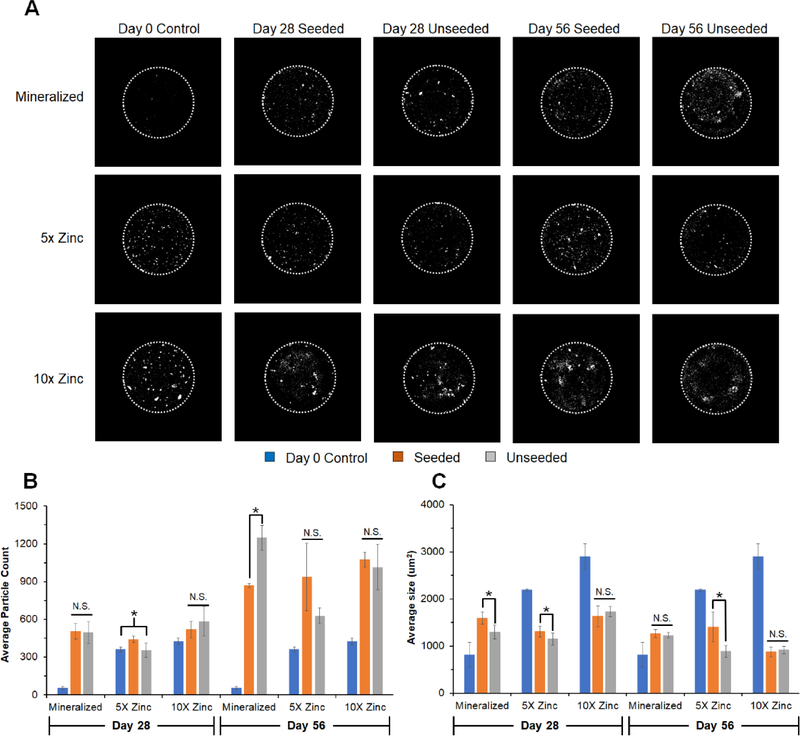
Figure 7: Micro-CT analysis of mineral remodeling over longe-term in vitro culture.
(A) Representative images (middle slice from each image stack) of mineralized, 1X Zinc, and 5X Zinc scaffolds along with unseeded controls. Diameter of scaffold is 6mm (dotted white line). (B) Average particle count representative of mineral nodules apparent within the scaffold. Overall, number of nodules increased from Day 28 to Day 56. All groups had significantly more particles than the unseeded Day 0 controls by Day 56. (C) Average particle size within scaffolds, showing mineralized scaffolds display larger particles compared to their unseeded Day 0 controls while the zinc functionalized scaffolds display significantly smaller particles compared to their unseeded Day 0 controls. *: significance at p<0.05 for indicated groups. N.S.: no significance between indicated groups.

