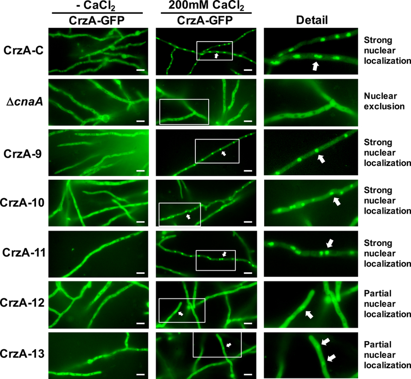
Figure 4. Calcium-stimulated nuclear translocation of mutant CrzA isoforms.

Conidia (104) of strains expressing GFP-labeled CrzA (CrzA-GFP) with native amino acid sequence (CrzA-C) or each mutant CrzA isoform CrzA-9 to CrzA-13 were inoculated into glass-bottomed Petri dishes containing liquid GMM and incubated overnight at 37°C. Hyphae were visualized microscopically prior to (CaCl -) and 15 minutes after addition of 200mM CaCl2 to the medium. A strain expressing native CrzA-GFP in a cnaA gene deletion background (ΔcnaA) was used as a negative control. Strong levels of calcium-stimulated nuclear translocation of CrzA were observed in strains CrzA-9, 10, and 11, but only partial nuclear translocation was observed in strains CrzA-12 and 13, while nuclear exclusion was observed in the ΔcnaA control. White scale bars represent 10μm distances. White arrows indicate examples of nuclear localization of CrzA-GFP where observed. CrzA-GFP = green fluorescence image. The enlarged detail images of nuclear and cytoplasmic localization of CrzA-GFP in the presence of 200mM CaCl2 are shown for clarity.
