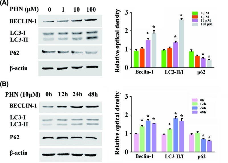
Figure 2. PHN triggered autophagy in HEp-2 cells.
(A) Proteins associated with autophagy were analyzed by Western blot with different concentrations of PHN (0, 1, 10 and 100 μM) treatment for 24 h. The expression level of BECLIN-1, LC3II/I and P62 and quantitation of the relative protein expression. (B) Proteins associated with autophagy were analyzed by Western blot with 10 μM PHN treatment for different times (0, 12, 24 and 48 h). The expression levels of BECLIN-1, LC3II/I ratio and P62 and quantitation of the relative protein expression. The experiment was repeated three times independently and data are expressed as mean ± standard error, *P<0.05.

