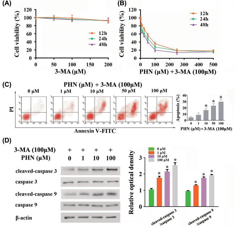
Figure 3. Effects of combining use of PHN and 3-MA in HEp-2 cells.
(A) Viability of HEp-2 cells treated with various concentrations of 3-MA for 12 h (orange), 24 h (green) and 48 h (purple) by CCK8 analysis. (B) Viability of HEp-2 cells treated with various concentrations of PHN and 100 μM 3-MA for 12 h (orange), 24 h (green) and 48 h (purple) by CCK8 analysis. (C) HEp-2 cells were stained with FITC-Annexin V/PI and analyzed by flow cytometry, with different concentrations of PHN (0, 1, 10, 50 and 100 μM) and 100 μM 3-MA treatment for 24 h. (D) Proteins associated with apoptosis were analyzed by Western blot. The expression level of cleaved-caspase 3, caspase 3, cleaved-caspase 9, caspase 9, β-actin and quantitation of cleaved-caspase 3/caspase 3 and cleaved-caspase 9/caspase 9 ratios. The experiment was repeated three times independently and data are expressed as mean ± standard error, *P<0.05.

