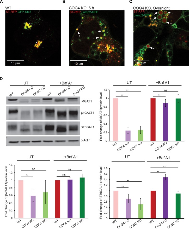
FIGURE 2.
Golgi enzymes partially co-localize with EELSs in COG KO cells and their stability is dependent on the activity of vacuolar ATPase. (A) ST-RFP is co-localized with Golgi marker GFP-STX5 in WT HEK 293T cells. (B) 6 h after transfection ST-RFP partially co-localizes with Lamp2-GFP positive EELSs in COG4 KO cells (Arrows point to EELSs, star indicates Golgi region). (C) After overnight expression in COG4 KO cells RFP fluorescence is seen within Lamp2 positive EELSs (arrows). (D) Western blot analysis of three Golgi enzymes in HEK293T cells before and after treatment of cells with 200 nm Baf A1 for 24 h. Bar graphs represent the fold change of Baf A1 treated vs. untreated protein levels. Error bars show SD for n = 3 (biological replicates). ∗∗p < 0.01.

