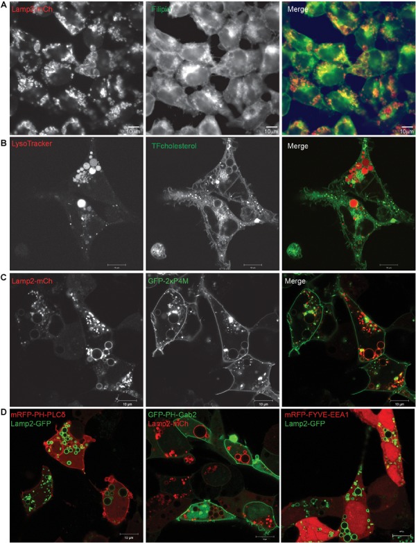
FIGURE 4.

Lipids present in EELSs’ membranes include PI4P and cholesterol. (A) Filipin staining in COG4 KO cells shows that EELSs’ membranes have cholesterol. (B) Presence of cholesterol was confirmed by TopFluor-cholesterol. EELSs are labeled by LysoTracker (C) Phosphatidylinositol PI4P, detected by the biosensor GFP-2×P4M colocalizes with Lamp2-mCh positive EELSs, (D) but, EELSs are negative for PI(4,5)P2, PI(3,4,5)P3, and PI3P detected by biosensors mRFP-PH-PLCδ, GFP-PH-Gab2, and mRFP-FYVE-EEA1, respectively. Scale bars are 10 μm.
