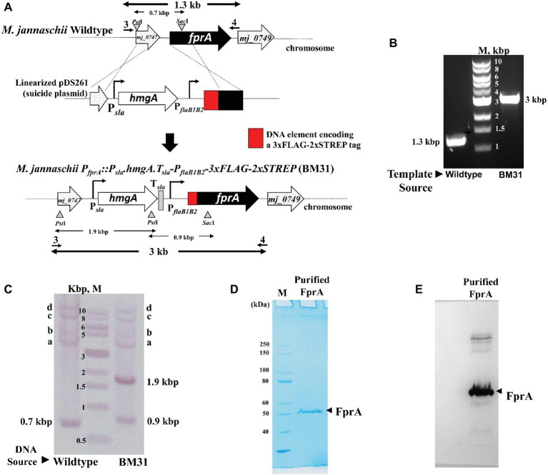
Figure 3.
Homologous expression of an affinity tagged protein driven by the flaB1B2 promoter in M. jannaschii. (A) Construction of M. jannaschii strain BM31 (PfprA::Psla.hmgA.Tsla.PflaB1B2-3xFLAG-Twin-Strep.fprA) expressing F420H2 oxidase (FprA) protein under the control of PflaB1B2, the promoter for the flagellin (flaB1B2) operon of M. jannaschii. The strain was constructed via double cross-over recombination between the upstream and coding regions of fprA (locus tag number, mj_0748) of the M. jannaschii chromosome and cloned homologous elements in a linearized form of pDS261, a suicide vector (Figure 1B). (B) PCR analysis of the genotype of M. jannaschii BM31. Primers 3 and 4, as shown in (A), were used for the amplification, and the respective sequences appear in Supplementary Table S1. The genomic regions targeted for PCR amplification are shown with bold double-sided arrows in (A). (C). Southern DNA hybridization analysis. Genomic DNA samples of wild-type and BM31 strains of M. jannaschii were digested with PstI and SacI. The suicide plasmid pDS261 was digested with HindIII and BamHI. The relevant restriction enzyme sites on the genome and suicide plasmid and the corresponding DNA fragments are shown with triangles/inverted triangles and double-sided arrows, respectively, in (A). A mixture of fragments resulting from the restriction enzyme digestion of pDS261 were labeled with Digoxigenin and used as hybridization probe. Observed hybridizing band, respective identity: 0.7, 0.9, and 1.9 kbp, shown in (A); a–c, PflaB1, fprA, and hmgA regions of M. jannaschii chromosome as shown in Supplementary Figure S2; d, partially digested high molecular mass DNA that hybridized with the DIG-labeled probes. (D) An SDS-PAGE profile of affinity purified Mj-FprA. Purified protein (0.5 μg) was analyzed on a 10% SDS-PAGE gel. The apparent molecular mass of Mj-FprA polypeptide was 54 kDa. M, unstained protein standard (New England Biolabs, Ipswich, MA). The masses of protein markers are shown next to the respective bands. (E) Western blot hybridization of Mj-FprA employing monoclonal anti-FLAG® M2 mouse antibody and visualized using an anti-mouse IgG (whole molecule) rabbit antibody conjugated with alkaline phosphatase and chromogenic alkaline phosphatase substrates, NBT and BCIP.

